.svg)
EEG with fNIRS, and the fight for space on the scalp
Neurovascular coupling, mobile brain-body imaging and hybrid BCIs all require electrical and haemodynamic measures from the same session. The constraint is physical: both sets of sensors compete for the same scalp. Which measure is primary decides the purchase.
.jpg)
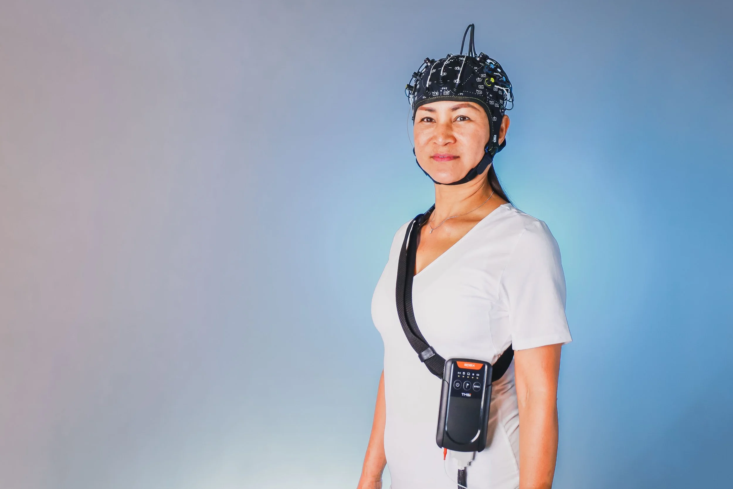
Neurovascular coupling, mobile brain-body imaging, and the literature on hybrid brain-computer interfaces that combine an electrophysiological feature with a haemodynamic one all require the same two measurements from the same session.
EEG resolves the timing of neural activity to the millisecond and localises it poorly. fNIRS localises cortical activation and resolves it over seconds, because the haemodynamic response is slow. Recorded together they answer when and where within one session, which is why EEG with fNIRS is the multimodal combination we install most frequently.
The constraint is physical, and it is the reason this guide exists. Both sets of sensors compete for the same scalp.
Why the two compete
An fNIRS channel is not a sensor. It is the volume of tissue sampled between one source optode and one detector optode, and the separation between that pair determines the depth at which the measurement is sensitive. At the standard adult separation of roughly 30 mm the sampled volume reaches cortex. Shorten the separation and it samples scalp instead. The separation is therefore set by the physics of photon migration, not by convenience.
EEG electrodes occupy the 10-20 positions. Optode pairs require specific separations at specific locations. Both sets of requirements apply to the same finite scalp area, so every electrode added removes a position an optode could have occupied, and the reverse.
That trade determines the design.
Decide which measure is primary
Before the purchase order, not after.
| Study design | Specification | Synchronisation |
| fNIRS supports an EEG study | g.SENSOR fNIRS on the amplifier already owned | None required, single device |
| fNIRS carries the finding | NIRSport2, NIRScout or Brite, electrodes fitted into the fNIRS cap | LSL and one trigger word |
| Both at high density | An integrated cap carrying both holder types, on a supported amplifier | Hardware TTL and LSL |
If EEG is primary
g.SENSOR fNIRS adds 8 fNIRS channels to a g.Nautilus, g.USBamp or g.HIamp already in the lab.
One amplifier records both modalities. No LSL, no trigger to distribute, no per-device offset to measure, because there is one clock and one converter.
g.tec state preparation for fNIRS plus 32 EEG channels at approximately 10 minutes with active electrodes. That figure determines how many participants are realistically run.
If fNIRS is primary
Specify NIRSport2 for mobile work, NIRScout for a laboratory bench, or an Artinis Brite.
Fit the EEG electrodes into the fNIRS cap. Artinis recommend this, and the reasoning is practical: their neoprene cap shields the detectors from ambient light and is rigid enough that optode positions remain stable.
Place electrodes on the 10-20 positions first and fit optodes between them. Both g.tec and Artinis give the same advice, because electrodes are the smaller and more tolerant of the two sensor types.
Where the optode template will not fit between the electrodes, Artinis state the options directly: divide the template into smaller sub-templates, or reduce the EEG channel count.
What an integrated cap is
At high density on both modalities, the solution is a cap manufactured to carry both sensor types in defined positions, rather than two caps or improvised mounting.
On the g.tec and NIRx combination this is the g.GAMMAcap. g.tec specify that it integrates g.SCARABEO active EEG electrodes and NIRx optodes within a single cap, supporting up to 64 EEG electrode holders alongside 32 optode holders, which accommodates 16 light sources and 16 detectors. Electrode holder rings and optode holder rings are moulded into the same cap at known positions, so placement is reproducible across sessions and participants rather than reconstructed each time.
The full specification is on the g.tec g.HIamp NIRx page, which documents up to 256 EEG channels with 16 sources and 16 detectors, dual-wavelength continuous-wave NIRS at 760 and 850 nm, and synchronisation over LSL and hardware TTL. A wireless equivalent is documented on the g.Nautilus NIRx page.
These caps exist only for particular amplifier and fNIRS combinations. Establish which combination supports one before assuming high density on both is available.
Where we would not spend the money
Where fNIRS exists to support an EEG study, prefrontal activation recorded alongside the ERPs, 8 channels on the amplifier already owned is the better specification than a second system. It costs less, it removes the synchronisation work entirely, and it prepares in around ten minutes.
Specify the dense fNIRS system when fNIRS carries the finding and EEG is the supporting measure.
What we would not do is supply 64-channel EEG and a dense optode array on the assumption that both run at full density. Without an integrated cap they do not, and that is normally discovered on the day of the first pilot.
Interference, and its direction
fNIRS is optical and does not disturb the EEG.
The electronics within the optodes can. Artinis report that reducing skin-electrode impedance minimises this interference, and that at sufficiently low impedance it is eliminated. The remedy is the electrode preparation the protocol should specify regardless, which makes this the least expensive problem in the guide.
Cap requirements
Dark, so ambient light does not reach the detectors. g.tec make the same point regarding cap material.
Rigid enough to hold optodes in fixed positions. An optode that shifts by a few millimetres is sampling a different volume of tissue.
Light. Two sensor sets on one head share the same movement artefacts.
Synchronisation
This is a smaller problem than with eye tracking. The haemodynamic response evolves over seconds while EEG resolves milliseconds, so sub-millisecond alignment is not the requirement.
Event markers must still be written into both recordings. A single device provides that inherently. Two systems require LSL and the same trigger word delivered to each, which is the architecture set out in eye tracking with EEG, and where the timing breaks.
For real-time and closed-loop work, g.tec run the EEG amplifier as the master device, because it has the higher sampling rate.
Short separation channels
A short channel uses a reduced source to detector separation to sample the extracerebral compartment, principally scalp blood flow, providing a regressor for removing that contribution from the long channels. NIRxWINGS2 records systemic physiology alongside.
Budget for these at specification stage. Adding them later requires reworking the optode layout, and the layout is what the electrodes were already competing for.
The parts
| Stage | Product |
| EEG amplifier | g.Nautilus Research, g.USBamp Research, g.HIamp |
| fNIRS on that amplifier | g.SENSOR fNIRS, 8 channels |
| Mobile fNIRS | NIRSport2, Artinis Brite |
| Laboratory fNIRS | NIRScout, Artinis Brite Ultra |
| Infants and children | Artinis BabyBrite, Brite Lite Frontal |
| Systemic physiology | NIRxWINGS2 |
| Analysis | NIRx Satori, Turbo-Satori for real time, Artinis OxySoft |
Where the choice between the two fNIRS platforms is still open, that comparison is on our fNIRS systems page and in the fNIRS buyer's guide.
NBT supplies g.tec, NIRx and Artinis in Israel, and installs them.
Send us the cortical area of interest, the EEG channel count required and the intended result, and we will specify which of the two routes applies. Which modality is carrying the finding, the electrical signal or the haemodynamic one?

NEED MORE INFORMATION ABOUT THIS PRODUCT?
Send us your emailFrequently Asked Questions
Can I record EEG and fNIRS from one device?
Yes, if EEG is the primary measure. g.SENSOR fNIRS adds 8 fNIRS channels to a g.Nautilus, g.USBamp or g.HIamp, so both signals are recorded by the same amplifier. One clock, no synchronisation to set up and nothing to measure afterwards.
How do optodes and electrodes share the same head?
Electrodes go on the 10-20 positions and optodes go in between, because electrodes are smaller and easier to place. An fNIRS channel is a pair of optodes about 30 mm apart, and that spacing sets the depth you are sensitive to, so it is not negotiable.
What is an integrated EEG and fNIRS cap?
A cap manufactured to carry both sensor types in defined positions. On the g.tec and NIRx combination this is the g.GAMMAcap, which g.tec specify as integrating g.SCARABEO active EEG electrodes and NIRx optodes in one cap, with up to 64 electrode holders alongside 32 optode holders for 16 sources and 16 detectors. Electrode and optode holder rings are moulded in at known positions, so placement is reproducible across sessions. These caps exist only for particular amplifier and fNIRS combinations, so confirm which combination supports one before assuming high density on both is available.
Do the two systems interfere with each other?
fNIRS is optical, so it does not disturb the EEG. The electronics in the optodes can. Artinis reports that lowering skin-electrode impedance minimises it, and that with low enough impedance it goes away, so the fix is the preparation you should be doing anyway.
Do I need short separation channels?
If the effect you are chasing is small, yes. Short channels measure the extracerebral signal, which is scalp blood flow rather than cortex, so you can regress it out of the long channels. Budget for them at the start, because retrofitting means rethinking the whole optode layout.
Still have questions?
Recent Blog Posts

.svg)
Advance Your Research
Contact NBT today for expert consultation on your neuroscience instrumentation needs.