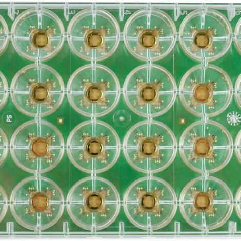
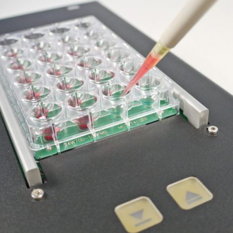
Overview
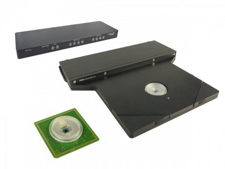
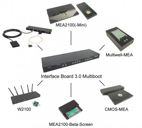
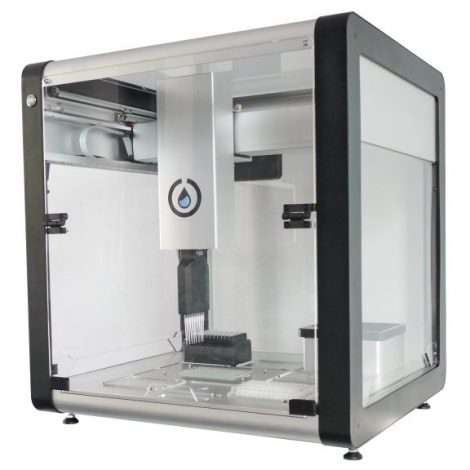
The CMOS-MEA5000-System consists of four components, which are all designed to be efficient and powerful, while fitting ideally on the lab bench and microscopes.
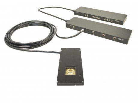
After opening the lid of the headstage, you just place the chip inside an. When the lid is closed, the contact pins are pressed on the pads on the chip and signals are transmitted. The headstage connects with only one cable (eSATA) to the interface board. Connection to the computer is done via USB 3.0. Summarizing, there is no need for many cables and the system is easy-to-use.
Please click on the arrows on the right to learn more about the single components.

The CMOS Chip
The chip is based on complementary metal oxide semiconductor (CMOS) technology, facilitating fast, high-resolution imaging of electrical activity.
The chip is equipped with a culture chamber to house your sample, while allowing the use of a microscope.
The recording sites are arranged in a 65×65 grid and have a 16µm or 32µm interelectrode distance (center to center). Between the recording electrodes, there is a grid of 32×32 bigger stimulation sites. Summarizing, you can record from 4225 electrodes and stimulate your sample at 1024 sites.
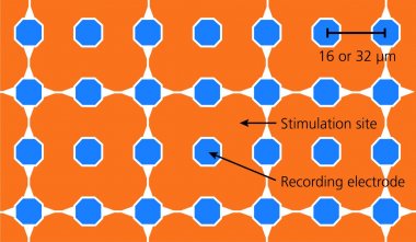 The high number of electrodes also results in a large recording area (1 mm² @ 16 µm; 4 mm² @ 32 µm). Thereby, you can see the signals from every single cell and even the signal propagation along an axon, while still getting an overview on your complete sample, e.g. a cell culture and see how the cells interact.
The advantages of the CMOS-MEA5000-System are:
- 4225 recording electrodes on an active chip
- 1024 dedicated stimulation sites
- 25 kHz sampling rate on all channels
- Outstanding signal quality
- Recordings at sub-cellular level
- Powerful software package CMOS-MEA-Control included (free updates available)
The chip is coated with planar oxide, similar to glass, enhancing the biocompatibility and biostability.

