.svg)
.svg)
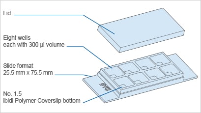
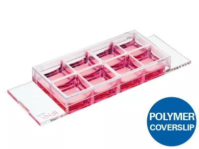
ibidi µ-Slide 8 Well
A chambered coverslip with 8 wells for cell culture, immunofluorescence, and high-end microscopy All-in-one 8 well chamber slide for cost-effective experiments—small number of cells and low volume of reagents needed In this microscopy slide, the cells are imaged on a No. 1.5 polymer coverslip bottom with the highest optical quality.


Technical Features
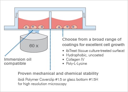
- Chambered coverslip with 8 independent wells and a non-removable polymer coverslip-bottom
- Now also available as a µ-Slide 8 Well high with extra high individual walls to keep cross contamination between wells as low as possible
- ibiTreat (tissue culture-treated) surface for optimal cell adhesion
- Imaging chamber slide with excellent optical quality for high-end microscopy
- Compatible with staining and fixation solutions
- Biocompatible plastic material—no glue, no leaking
- Also available as an adhesive version without a bottom: sticky-Slide 8 Well
- Also available with a Glass Coverslip Bottom: µ-Slide 8 Well Glass Bottom for special microscopic applications
- Additional version available with a 500 µm grid: µ-Slide 8 Well Grid-500
.webp)
The Principle of the µ-Slide 8 Well
The Coverslip Bottom
The µ-Slide 8 Well comes with a thin ibidi Polymer Coverslip Bottom that has the highest optical quality (comparable to glass) and is ideally suitable for high-resolution microscopy. It is also available as a sticky version without any bottom, or and as an option with a Glass Coverslip Bottom for special microscopic applications.
Find more information and technical details about the coverslip bottom of the ibidi chambers
Application Examples
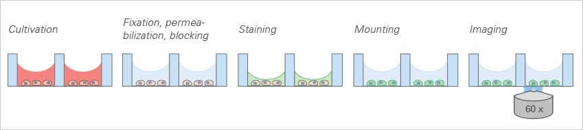
Immunofluorescence
The ibidi µ-Slide 8 Well allows for standard immunofluorescence protocols to be employed without the use of coverslips in an all-in-one chamber. All steps (e.g., cell cultivation, fixation, staining, and imaging) are carried out in the open well geometry. After staining, the sample can be observed through the coverslip bottom using high-resolution microscopy.
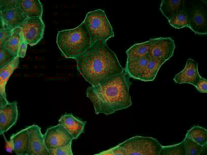
Fluorescence microscopy of MDCK cells. Mitochondria (MitoTracker, red), Actin cytoskeleton (Phalloidin, green), nuclei (DAPI, blue).
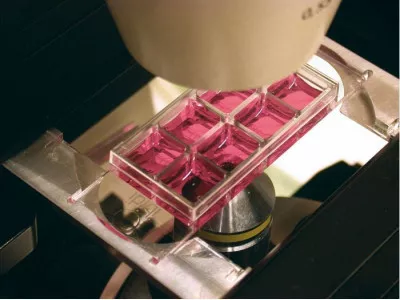
Live Cell Imaging
The µ-Slide 8 Well enables high-resolution live cell imaging using different brightfield and fluorescence techniques. Here, live cell microscopy was performed using the µ-Slide 8 Well in the ibidi Heating System, Universal Fit, for 1 Chamber on a Nikon Eclipse TIE inverted microscope.
Live cell imaging in a µ-Slide 8 Well using transmitted light.
F-actin Visualization in Living Cells Using LifeAct-TagGFP2 Protein
Live cell imaging of F-actin in Rat1 fibroblasts after LifeAct-TagGFP2 Protein transfer (30 µg/ml, 3 minutes) using the µ-Slide 8 Well.
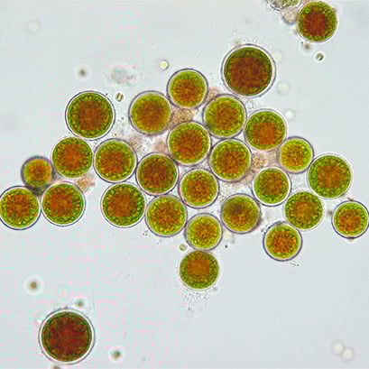
Live Cell Imaging of Algae
Brightfield microscopy of the green freshwater algae Haematococcus pluvialis in a µ-Slide 8 Well. The cells are in an immature cyst state with developing centers, filled with the strong antioxidant astaxanthin (red), and surrounded by Chlorophyll from the chloroplasts (green). 40x objective lens.
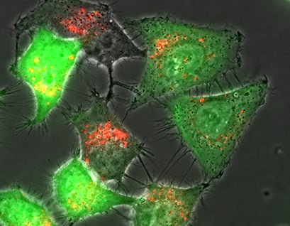
Live Cell Imaging of Fibroblasts After Transfection
Live cell imaging on an inverted widefield fluorescence microscope. Rat fibroblast cells 24 hours after transfection with pCMV-eGFP. Red: Rhodamine-labelled fluorescent lipoplexes, Green: eGFP fluorescence. 60x objective lens.
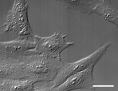
DIC Microscopy of Mammalian Cells
Differential interference contrast (DIC) microscopy of mammalian cell culture (fibroblasts) using the µ-Slide 8 Well and the DIC lid for µ-Slides. 63x objective lens.
Cell Cultivation and Imaging
Explore All ProductsFluorescence Imaging
Explore All Productsibidi Labware Publications
Comprehensive collection of peer-reviewed research publications demonstrating the use of ibidi labware including µ-Slides, µ-Dishes, µ-Plates, and specialized microfluidic devices for cell biology, neurobiology, vascular biology, and cancer research.
What is the µ-Slide 8 Well used for?
Eight-chamber slide for immunofluorescence, live-cell imaging, and multi-step assays. Individual isolation walls and #1.5 Polymer or Glass Coverslip bottom deliver cost-effective parallel microscopy across standard inverted platforms.
Is it compatible with high-resolution microscopy?
Yes — features ibidi's premium #1.5 polymer coverslip bottom, ideal for widefield fluorescence, DIC, phase contrast, and confocal imaging. For super-resolution and TIRF, choose the Glass Bottom variant.
Is the µ-Slide 8 Well sterile and ready to use?
Yes. All ibidi labware ships sterilized and individually wrapped, ready for direct cell seeding without additional preparation.
What surface treatment options are available for the µ-Slide 8 Well?
ibidi labware is offered in multiple surface treatments including ibiTreat (tissue-culture treated), Bioinert, hydrophobic, and uncoated — selected to match adherent or suspension cell requirements.
Is the µ-Slide 8 Well available with local Israeli support?
Yes. NBT Ltd is the exclusive ibidi distributor in Israel, providing application training, installation guidance, and full local technical support for the µ-Slide 8 Well.
Still have questions?
Request for Quote
Please fill in as much details as possible and we will take care of your request as soon as possible
Request for Quote
Please fill in as much details as possible and we will take care of your request as soon as possible