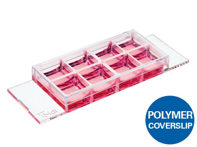
Applications
- Cultivation and microscopy of cell cultures
- Transfection assays
- Fluorescence microscopy of both living and fixed cells
- Live cell imaging over extended time periods
- Suitable for DIC, when used with a DIC lid
Want to know if you should use a glass or a polymer bottom for your application? Find out here.
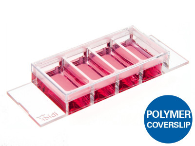
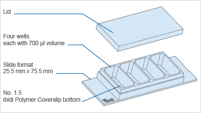
Specifications
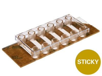
| Outer dimensions (w x l) | 25.5 x 75.5 mm² |
| Number of wells | 4 |
| Dimensions of wells (w x l x h) |
21.6 x 11.4 x 9.3 mm³ |
| Volume per well | 700 µl |
| Total height with / without lid | 10.8 / 9.5 mm |
| Growth area per well | 2.5 cm² |
| Coating area per well | 4.1 cm² |
| Bottom: ibidi Polymer Coverslip | |
Technical Features
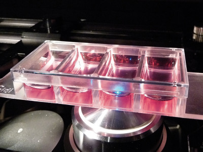
- Open µ-Slide (chambered coverslip) with 4 independent wells
- Excellent optical quality imaging chamber for high-end microscopy
- Compatible with staining and fixation
- ibiTreat surface for optimal cell adhesion
- Made of biocompatible plastic material – no glue and no leaking
- Minimal mirror effect on the µ-Slide walls for light sheet microscopy
- Also available with a glass bottom
- Available with a non-adhesive Bioinert surface: µ-Slide 4 Well Bioinert, specifically suitable for 3D applications, such as spheroids and suspension cells
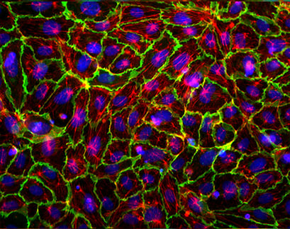
Experimental Example
Immunofluorescence of Human Umbilical Vein Endothelial Cells (HUVEC) in a µ-Slide 4 Well
Blue: Nucleus (DAPI)
Green: VE-Cadherin (Alexa555)
Red: F-Actin (Alexa488)
Meniscus-Free Phase Contrast Microscopy
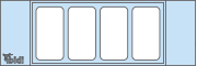
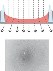
| µ-Slide 4 Well
|
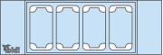
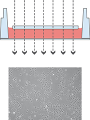
µ-Slide 4 Well Ph+
|
| Top View
|
|
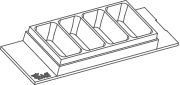
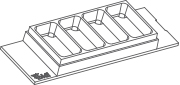
The µ-Slide 4 Well Ph+ (Phase Contrast +) is designed for excellent optical quality, especially for cell culture when normal phase contrast microscopy is being used. Opposed to the classic µ-Slide 4 Well, the Ph+ version provides a special intermediate plate in the center of the well. This plate flattens the meniscus that disturbs the phase contrast effect in normal open wells. Openings near the corners provide access to the wells for easy filling and aspiration of liquids.
This innovative technique supports meniscus-free phase contrast microscopy in a very convenient manner.
Working with the µ-Slide 4 Well Ph+ diminishes the meniscus and increases the area of well contrasted cells
 |
 |
The illustration on the left shows the perturbing effect of a meniscus. Light is refracted on the air-water-interface, leading to poor contrast in microscopy. Only the small center part exhibits satisfying phase contrast.
Working with the µ-Slide 4 Well Ph+ diminishes the meniscus and increases the area of nicely contrasted cells. This nice contrast is due to the parallel beam