Applications
- Cultivation and high-resolution microscopy of cells
- Fluorescence microscopy of living and fixed cells
- Immunofluorescence staining
- Live cell imaging over extended time periods
- Transfection assays
- Differential interference contrast (DIC) when using a DIC lid
Want to know if you should use a glass or a polymer bottom for your application? Find out here.
Specifications
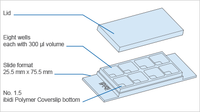
| Outer dimensions (w x l) | 25.5 x 75.5 mm² |
| Number of wells | 8 |
| Dimensions of wells (w x l x h) | 9.4 x 10.7 x 6.8 mm³ |
| Volume per well | 300 µl |
| Total height with lid | 8 mm |
| Growth area per well | 1.0 cm² |
| Coating area per well | 2.20 cm² |
| Bottom: ibidi Polymer Coverslip | |
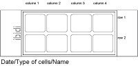
Define and print your experimental setup
Technical Features
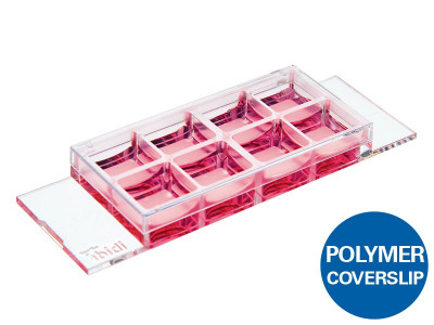
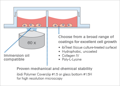
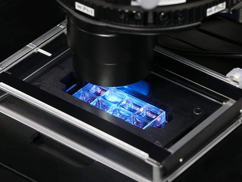
- Chambered coverslip with 8 independent wells and a non-removable polymer coverslip-bottom
- Now also available as a µ-Slide 8 Well high with extra high individual walls to keep cross contamination between wells as low as possible
- ibiTreat (tissue culture-treated) surface for optimal cell adhesion
- Imaging chamber slide with excellent optical quality for high-end microscopy
- Compatible with staining and fixation solutions
- Biocompatible plastic material—no glue, no leaking
- Also available as an adhesive version without a bottom: sticky-Slide 8 Well
- Also available with a Glass Coverslip Bottom: µ-Slide 8 Well Glass Bottom for special microscopic applications
- Additional version available with a 500 µm grid: µ-Slide 8 Well Grid-500
The Principle of the µ-Slide 8 Well
The Coverslip Bottom
The µ-Slide 8 Well comes with a thin ibidi Polymer Coverslip Bottom that has the highest optical quality (comparable to glass) and is ideally suitable for high-resolution microscopy. It is also available as a sticky version without any bottom, or and as an option with a Glass Coverslip Bottom for special microscopic applications.
Find more information and technical details about the coverslip bottom of the ibidi chambers here.
The ibiTreat Surface
ibiTreat (tissue culture-treated) is our most recommended surface modification, because almost all adherent cells grow well on it without the need for any additional coating.
Find more information about the different surfaces of the ibidi chambers here.
Application Examples
Immunofluorescence
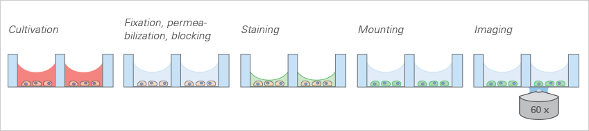
The ibidi µ-Slide 8 Well allows for standard immunofluorescence protocols to be employed without the use of coverslips in an all-in-one chamber. All steps (e.g., cell cultivation, fixation, staining, and imaging) are carried out in the open well geometry. After staining, the sample can be observed through the coverslip bottom using high-resolution microscopy.
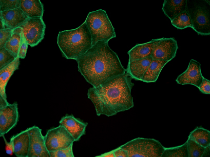
Fluorescence microscopy of MDCK cells. Mitochondria (MitoTracker, red), Actin cytoskeleton (Phalloidin, green), nuclei (DAPI, blue).
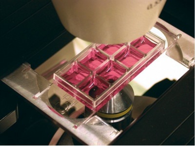
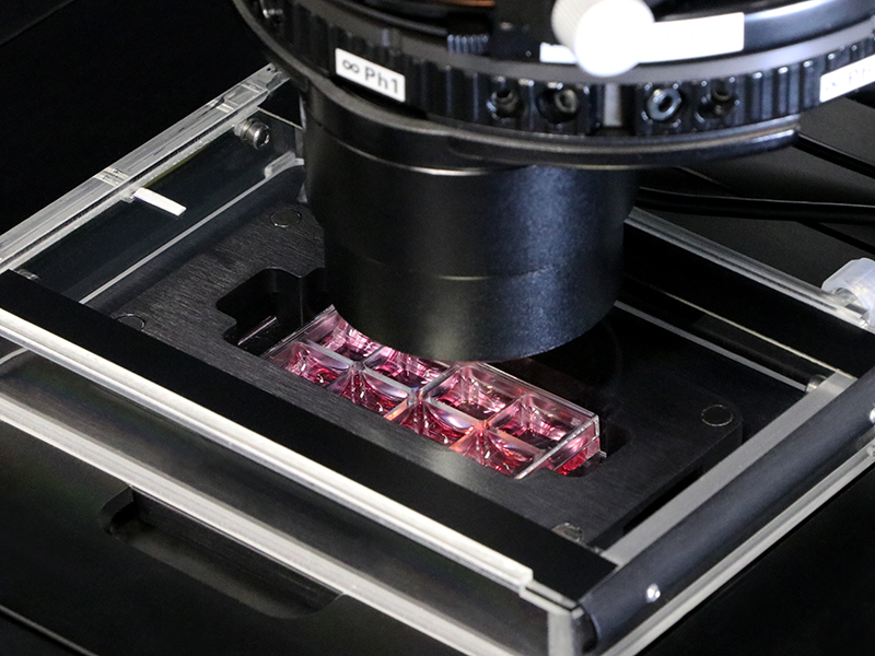
Live Cell Imaging
The µ-Slide 8 Well enables high-resolution live cell imaging using different brightfield and fluorescence techniques. Here, live cell microscopy was performed using the µ-Slide 8 Well in the ibidi Heating System, Universal Fit, for 1 Chamber on a Nikon Eclipse TIE inverted microscope.
Live cell imaging in a µ-Slide 8 Well using transmitted light.
Live cell imaging in a µ-Slide 8 Well using fluorescence.

F-actin Visualization in Living Cells Using LifeAct-TagGFP2 Protein
Live cell imaging of F-actin in Rat1 fibroblasts after LifeAct-TagGFP2 Protein transfer (30 µg/ml, 3 minutes) using the µ-Slide 8 Well.
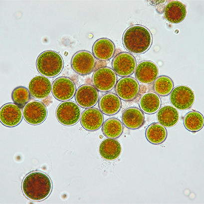
Live Cell Imaging of Algae
Brightfield microscopy of the green freshwater algae Haematococcus pluvialis in a µ-Slide 8 Well. The cells are in an immature cyst state with developing centers, filled with the strong antioxidant astaxanthin (red), and surrounded by Chlorophyll from the chloroplasts (green). 40x objective lens.
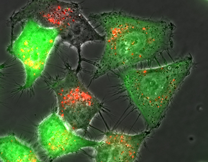
Live Cell Imaging of Fibroblasts After Transfection
Live cell imaging on an inverted widefield fluorescence microscope. Rat fibroblast cells 24 hours after transfection with pCMV-eGFP. Red: Rhodamine-labelled fluorescent lipoplexes, Green: eGFP fluorescence. 60x objective lens.
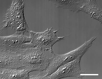
DIC Microscopy of Mammalian Cells
Differential interference contrast (DIC) microscopy of mammalian cell culture (fibroblasts) using the µ-Slide 8 Well and the DIC lid for µ-Slides. 63x objective lens.