Applications
- Cultivation and high-resolution microscopy of cells
- TIRF and single molecule applications of living and fixed cells
- Super-resolution microscopy
- Immunofluorescence staining and fluorescence microscopy of living and fixed cells
- Live cell imaging over extended time periods
- Transfection assays
- Differential interference contrast (DIC) microscopy when used with a DIC lid
Want to know if you should use a glass or a polymer bottom for your application? Find out here.
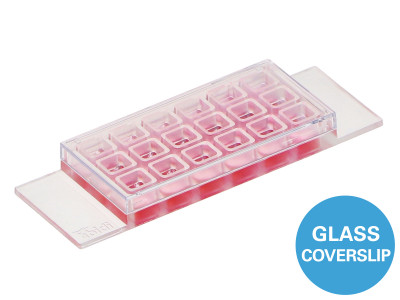
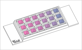
Specifications
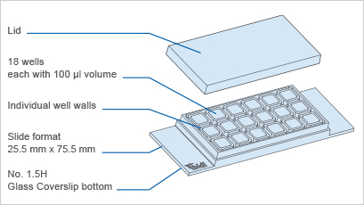
| Outer dimensions (w x l) | 25.5 x 75.5 mm² |
| Number of wells | 18 |
| Dimensions of wells (w x l x h) | 5.7 x 6.1 x 6.8 mm³ |
| Volume per well | 100 µl |
| Height with/without lid | 8.2/6.8 mm |
| Growth area per well | 0.34 cm² |
| Coating area per well | 1.15 cm² |
| Bottom: Glass coverslip No. 1.5H, selected quality, 170 µm +/- 5 µm | |
Technical Drawing
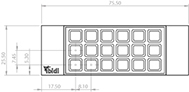
Technical drawings and details are available in the Instructions (PDF).
Technical Features
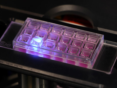
- Chambered glass coverslip with 18 independent wells
- Bottom made from D 263 M Schott glass, No. 1.5H (170 +/- 5 µm)
- May require coating to promote cell attachment
- Imaging chamber slide with excellent optical quality for high-end microscopy
- Closely fitting lid for low evaporation
- Individual well walls for minimizing well-to-well crosstalk and contaminations
- Compatible with staining and fixation solutions
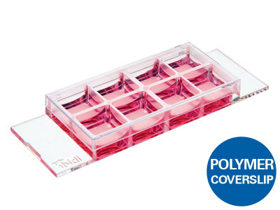
- Also available with an ibidi Polymer Coverslip Bottom for superior cell growth: µ-Slide 18 Well
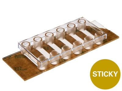
- Also available as an adhesive version without a bottom: sticky-Slide 18 Well
Application Examples
Multiple conditions on one µ-Slide: For immunofluorescence stainings, toxicological screenings, cell surface coatings.
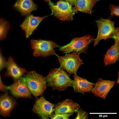
Fluorescence microscopy of rat fibroblasts (Rat1) in a µ-Slide 18 Well Glass Bottom. Red: alpha-tubulin, green: F-actin, stained with LifeAct-TagGFP2 Protein; blue: nuclei (ibidi Mounting Medium with DAPI). 60x objective lens, oil immersion.