.svg)
Lower Limb Prosthetics
Myoelectric-controlled lower limb prostheses use EMG signals to actuate movement, but assumptions about neural activation between residual and intact limbs may need revision. New research reveals that amputation alters motor unit properties in ways that directly impact prosthetic control models.

Key Takeaways
- Myoelectric control uses EMG signals to control actuation of an external device such as a prosthetic. Motor unit (MU) information from the amputated limb can provide additional inputs to the controller algorithm.
- It was a common assumption that neural activation between the residual limb and intact limb were the same when creating a model of muscle recruitment.
- Research shows that alterations of MU properties exist within the residual limb, holding implications for prosthesis control — myoelectric models may need reparameterization to account for these neuromuscular changes.
What are Myoelectric-Controlled Lower Limb Prostheses?
Myoelectric control is the use of EMG signals to control actuation of an external device, typically a prosthetic. EMG signals are input into a processor and classifier that predicts the user's intended movement, translating intent into an actuated motion. The main goal is to activate the prosthesis in a naturalistic fashion — as an intact limb would.
Incorporation of Motor Unit Data
With the advancement of EMG sensors and decomposition algorithms, researchers can now isolate individual motor unit (MU) information. MU information provides additional insights to the controller for a better representation of intended movement. However, recent research reveals that after amputation, damage to the peripheral neuromuscular system disrupts afferent feedback and alters the internal representation of central motor control and EMG activity.
How EMG Was Used
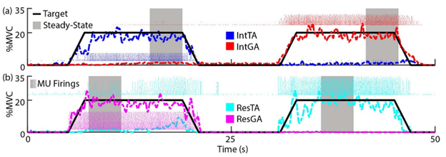
EMG was recorded on the tibialis anterior (TA) and lateral gastrocnemius (GA) of both residual and intact limbs. sEMG signals were acquired and decomposed through the Delsys Galileo sensor: four-pin array electrodes. The 5mm interelectrode distance minimized crosstalk between muscles. The raw EMG signal was decomposed by Delsys NeuroMap software to obtain the MU action potentials and timing of MU firings.
What Was Found
In intact muscles, motor unit amplitudes usually increase as the recruitment timeline increases. However, the residual tibialis anterior (TA) exhibited less correlation between size and recruitment timeline. Regarding the onion-skin scheme, intact limbs typically display a negative correlation between motor unit mean firing rates and recruitment threshold — in the residual TA, that negative correlation is flattened. These changes show that neuromuscular control is much slower in the residual TA than in the intact TA.
Future Impact
The evidence convinces researchers to reject the assumption that EMG generation properties between residual and intact muscles are similar. For future research in EMG modeling, default parameterization of MU pool organization should not be assumed for residual muscles. The type of muscles and the alteration of correlative relationships between the size principle, onion-skin model, and rate-size association need to be taken into account for better representing motor intent in musculoskeletal model-based myoelectric controllers.

Shall we chat about this topic?
Contact usRecent Blog Posts


%3Aquality(100).webp)

Advance Your Research
Contact NBT today for expert consultation on your neuroscience instrumentation needs.