.svg)
From Lab to Life: Transforming Movement Studies with Dynamic TMS and EMG
Cardiff University researchers combine TMS and Delsys EMG during dynamic walking tasks to reveal how the brain and spinal cord coordinate muscle activity in real-world movement — with direct applications in fall prevention and rehabilitation.

Key Takeaways
- During traditional TMS research, most studies take place with participants sitting still — far from the complex movements we make every day.
- EMG recordings capture the muscles' immediate responses to TMS pulses, offering a direct window into brain-muscle communication.
- An innovative system at Cardiff University combines TMS and EMG during dynamic activities, bridging the gap between lab findings and real-world movement.
Current Neurophysiology Research
The brain plays a central role in controlling the muscles in everything we do. Whilst biomechanical research into muscle activation is common, neurophysiology research into how the brain connects to the muscles is less so. Understanding this connection is imperative to learn what, from a neural perspective, has led to a biomechanical change.
Dr. Jennifer Davies, Senior Lecturer at Cardiff University, uses Transcranial Magnetic Stimulation (TMS) — a non-invasive technique that uses magnetic fields to stimulate nerve cells in the brain — in conjunction with Delsys EMG technology. EMG is critical because muscle activity is the readout of how responsive the neural system is. TMS cannot tell us anything about the neural control of muscle without it.
"We're really good at describing changes that happen biomechanically. But if we're trying to target something like that from a rehabilitation perspective, we still don't know from a neural perspective what has changed to cause this biomechanical change." — Dr. Jennifer Davies, Cardiff University
Integrating TMS and EMG into a Dynamic Testing Environment
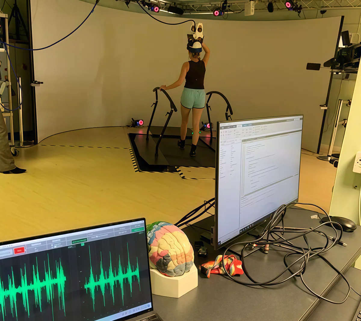
Cardiff University, in collaboration with Magstim and Cardiff University Brain Research Imaging Centre (CUBRIC), developed a novel method of delivering brain stimulation during walking. The TMS coil is held in a helmet supported by springs, maintaining precise coil position during movement. The helmet integrates with a MOTEK Grail treadmill system, allowing biomechanics to be tested under different conditions such as incline, decline, and gait perturbations.
Delsys Trigno Avanti sensors provide the critical real-time EMG readout of how the nervous system responds to stimulation across a wide number of scenarios. TMS is delivered at precise moments during the gait cycle — heel strike, mid-stance, and toe-off — allowing Jen to assess how the nervous system responds dynamically, rather than in static conditions.
Implications of Dynamic TMS Outside the Laboratory
Research is now underway to help understand why dynamic biomechanical changes occur at a neural level. In a published study (Huiberts, Bruijn and Davies, 2025), the team investigated how corticospinal excitability responded to mediolateral gait instability during walking. They found increased corticospinal excitability during unpredictable lateral destabilisations, and notably that the brain is preparing for instability even before the muscles react — providing key insights for fall prevention strategies and rehabilitation.
By combining TMS, EMG, and dynamic environments, Jen and her colleagues are transforming how we study movement and bringing laboratory science closer to real-world applications across biomechanical, clinical, and neurophysiological fields.
References
Huiberts, R., Bruijn, S., Davies, J. (2025). Corticospinal Excitability in Response to Mediolateral Gait Instability.

Shall we chat about this topic?
Contact usRecent Blog Posts


Advance Your Research
Contact NBT today for expert consultation on your neuroscience instrumentation needs.