When to use a Compresstome vs. Cryostat

With all sorts of tissue slice models on the market, how do you know when to use what type of instrument? In this article, we will review the most common types of tissue slicers, and discuss the best research and clinical applications of each type of equipment.

Main Types of Tissue Slicers:

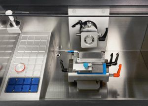
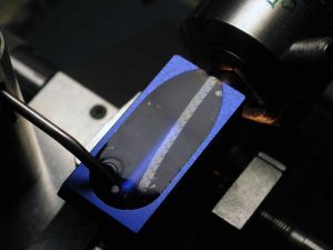
A tissue slicer is used to do exactly as its name implies: to cut tissue samples! Making tissue slices of consistent and controllable thicknesses is important because it allows for biologists and clinicians to obtain a standardized sample for further processing and experiments. The main types of tissues slicers include the following:

Vibrating microtomes: This type of slicer operates by cutting using a vibrating blade at high frequencies. This allows for cutting of live tissue (although fixed tissue can also be cut). The Compresstome is a type of vibrating microtome, as is the Vibratome by Leica.

Rotary microtomes: This type of tissue slicer operates with a staged rotary action, where the knife is fixed in a horizontal position, and the specimen holder rotates and cutting is done as part of the rotary motion. These slicers can be used on paraffin wax-embedded samples, which are very hard materials.
Cryostats: Also known as a “cryomicrotome,” cryostats are used to cut frozen samples. Typically, tissue samples are embedded in OCT media, which when frozen allows the hardness of the sample to be greatly increased. These slicers are used for both academic research and for doing biopsy analyses during surgery.
Ultramicrotome: These slicers allow preparation of extremely thin slices, and functions similarly to a rotary microtome. However, there is very careful mechanical construction which allows for very fine control of the cutting thickness. These slicers are used to obtain slices for transmission electron microscopy (TEM) and serial block-face scanning electron microscopy (SBFSEM).
Here’s a summary of the applications of when and what to use for specific tissue slicer types.