.svg)
.svg)
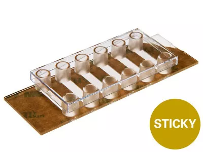
ibidi sticky-Slide VI 0.4
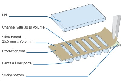
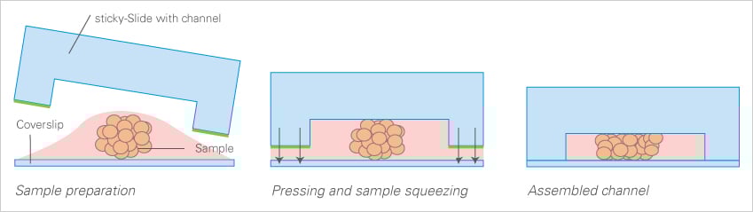
A bottomless 6 channel slide used for cell culture applications with a self-adhesive underside to which own substrates can be mounted Versatile.
Applications:
- Running multiple immunofluorescence assays and live cell imaging
- Real-time imaging under either static or flow conditions
- Inserting materials or tissue into perfusion channels
- Running of shear stress experiments on any substrate
- Usable with a range of bottom materials, such as plastic sheets, silicon chips, and glass slides
.webp)

Technical Features:
- Bottomless slide
- Self-adhesive underside
- Biocompatible adhesive that has been cell culture tested
- Adheres to all flat surfaces, even wet surfaces
- Female Luer adapters for connection to tubing and pump systems
- Sterile packaging
- Suitable coverslips available
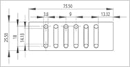
- Specifications identical to those of the µ-Slide VI 0.4, except that it has no bottom
Cell Cultivation and Imaging
Explore All ProductsCell Culture Under Flow
Explore All ProductsFluorescence Imaging
Explore All Productsibidi Labware Publications
Comprehensive collection of peer-reviewed research publications demonstrating the use of ibidi labware including µ-Slides, µ-Dishes, µ-Plates, and specialized microfluidic devices for cell biology, neurobiology, vascular biology, and cancer research.
What is the sticky-Slide VI 0.4 used for?
Six-channel adhesive slide bonded to the user's substrate of choice — coverslip, gel, or membrane. Identical channel geometry to µ-Slide VI 0.4 with full flexibility for custom surface chemistry, patterning, or specialized substrates.
Is it compatible with high-resolution microscopy?
Yes — features ibidi's premium #1.5 polymer coverslip bottom, ideal for widefield fluorescence, DIC, phase contrast, and confocal imaging. For super-resolution and TIRF, choose the Glass Bottom variant.
Is the sticky-Slide VI 0.4 sterile and ready to use?
Yes. All ibidi labware ships sterilized and individually wrapped, ready for direct cell seeding without additional preparation.
What surface treatment options are available for the sticky-Slide VI 0.4?
ibidi labware is offered in multiple surface treatments including ibiTreat (tissue-culture treated), Bioinert, hydrophobic, and uncoated — selected to match adherent or suspension cell requirements.
Is the sticky-Slide VI 0.4 available with local Israeli support?
Yes. NBT Ltd is the exclusive ibidi distributor in Israel, providing application training, installation guidance, and full local technical support for the sticky-Slide VI 0.4.
Still have questions?
Request for Quote
Please fill in as much details as possible and we will take care of your request as soon as possible
Request for Quote
Please fill in as much details as possible and we will take care of your request as soon as possible