.svg)
.svg)
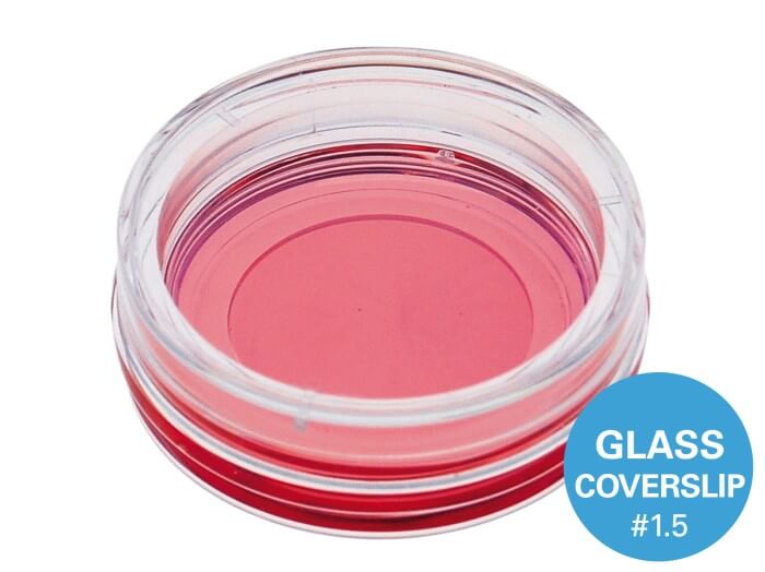
µ-Dish 35 mm, high Glass Bottom
A 35 mm imaging dish with a glass bottom for use in TIRF, single molecule and super-resolution microscopy applications
Technical Features
- Standard format imaging dish with a 35 mm diameter for tissue culture
- Bottom made from D 263 M Schott glass with a thickness of 170 µm +/- 5 µm
- May require coating to promote cell attachment
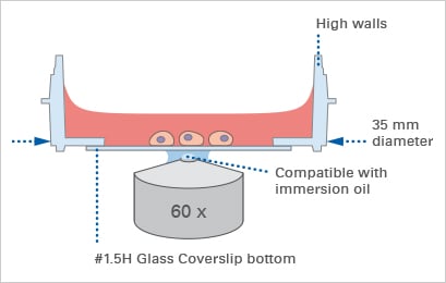
- High walls with a standard height for easy handling
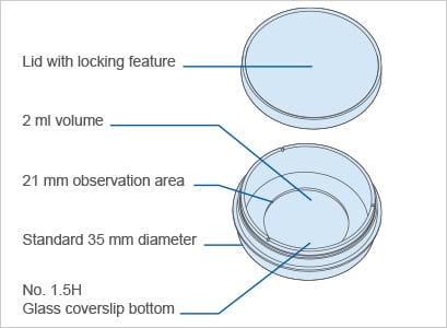
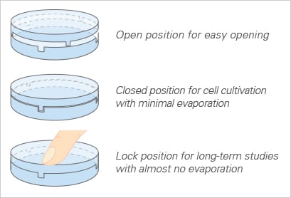
- Lid with locking feature for minimal evaporation
- Rim for easy opening
- No autofluorescence
- Fully biocompatible materials
- Available as a Bulk Box with 400 individually packed µ-Dishes per box
- Available with a #1.5 ibidi Polymer Coverslip Bottom for optimized adhesion
- Available with a 50 and 500 µm grid for cell counting
Test our everyday solution for cost-effective high-throughput experiments: Glass Bottom Dish 35 mm
The Coverslip Bottom
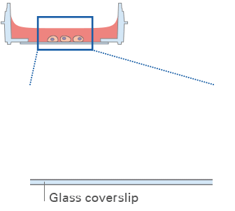
The µ-Dish 35 mm, high Glass Bottom comes with a thin #1.5H Glass Coverslip Bottom made from D 263 M Schott borosilicate glass that has the highest optical quality. ibidi developed these glass surfaces specifically for TIRF, super-resolution microscopy, and single molecule microscopy.
Lid with Locking Feature for Minimized Evaporation
All ibidi µ-Dishes are equipped with the special lid-locking feature. The locking position minimizes evaporation, providing excellent conditions for long-term studies in a non-humidified environment. Gas exchange during cell culture is maintained thanks to the gas-permeable plastic material.
.webp)

Technical Specifications
| Dish Diameter | 35 mm |
| Volume | 2 ml |
| Growth Area | 3.5 cm² |
| Coating Area (400 µl) | 4.1 cm² |
| Observation Area Diameter | 21 mm |
| Height (with lid / without lid) | 14 mm / 12 mm |
| Bottom Material | Glass Coverslip No. 1.5H, D 263 M Schott borosilicate glass, 170 µm ± 5 µm |
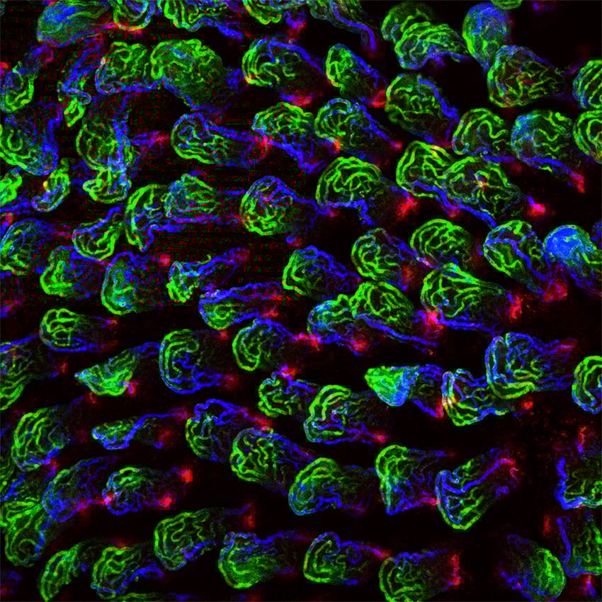
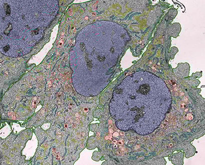
Applications
- Cultivation and high-resolution microscopy of cells
- Total Internal Reflection Fluorescence (TIRF) and single molecule applications
- Super-Resolution Microscopy (STED, SIM, (F)PALM, (d)STORM) and Fluorescence Correlation Spectroscopy (FCS)
- Immunofluorescence staining of living and fixed cells
- Widefield and confocal fluorescence microscopy
- Live cell imaging over extended time periods
- Transfection assays
- Differential Interference Contrast (DIC) microscopy
- Cell location and counting (gridded version available)
Cell Cultivation and Imaging
Explore All ProductsFluorescence Imaging
Explore All Productsibidi Labware Publications
Comprehensive collection of peer-reviewed research publications demonstrating the use of ibidi labware including µ-Slides, µ-Dishes, µ-Plates, and specialized microfluidic devices for cell biology, neurobiology, vascular biology, and cancer research.
What is the µ-Dish 35 mm, high Glass Bottom used for?
35 mm cell culture dish with a high-quality #1.5H glass coverslip bottom for high-resolution live-cell imaging. The high-walled design accommodates larger media volumes for long-term experiments while maintaining excellent optical access.
Is the µ-Dish 35 mm, high Glass Bottom compatible with high-resolution microscopy?
Yes. The µ-Dish 35 mm, high Glass Bottom features ibidi's premium #1.5 polymer or glass coverslip-equivalent bottom, compatible with confocal, fluorescence, TIRF, and high-NA oil-immersion objectives on standard inverted microscopes.
Is the µ-Dish 35 mm, high Glass Bottom sterile and ready to use?
Yes. All ibidi labware ships sterilized and individually wrapped, ready for direct cell seeding without additional preparation.
What surface treatment options are available for the µ-Dish 35 mm, high Glass Bottom?
ibidi labware is offered in multiple surface treatments including ibiTreat (tissue-culture treated), Bioinert, hydrophobic, and uncoated — selected to match adherent or suspension cell requirements.
Is the µ-Dish 35 mm, high Glass Bottom available with local Israeli support?
Yes. NBT Ltd is the exclusive ibidi distributor in Israel, providing application training, installation guidance, and full local technical support for the µ-Dish 35 mm, high Glass Bottom.
Still have questions?
Request for Quote
Please fill in as much details as possible and we will take care of your request as soon as possible
Request for Quote
Please fill in as much details as possible and we will take care of your request as soon as possible