

Skinscanner
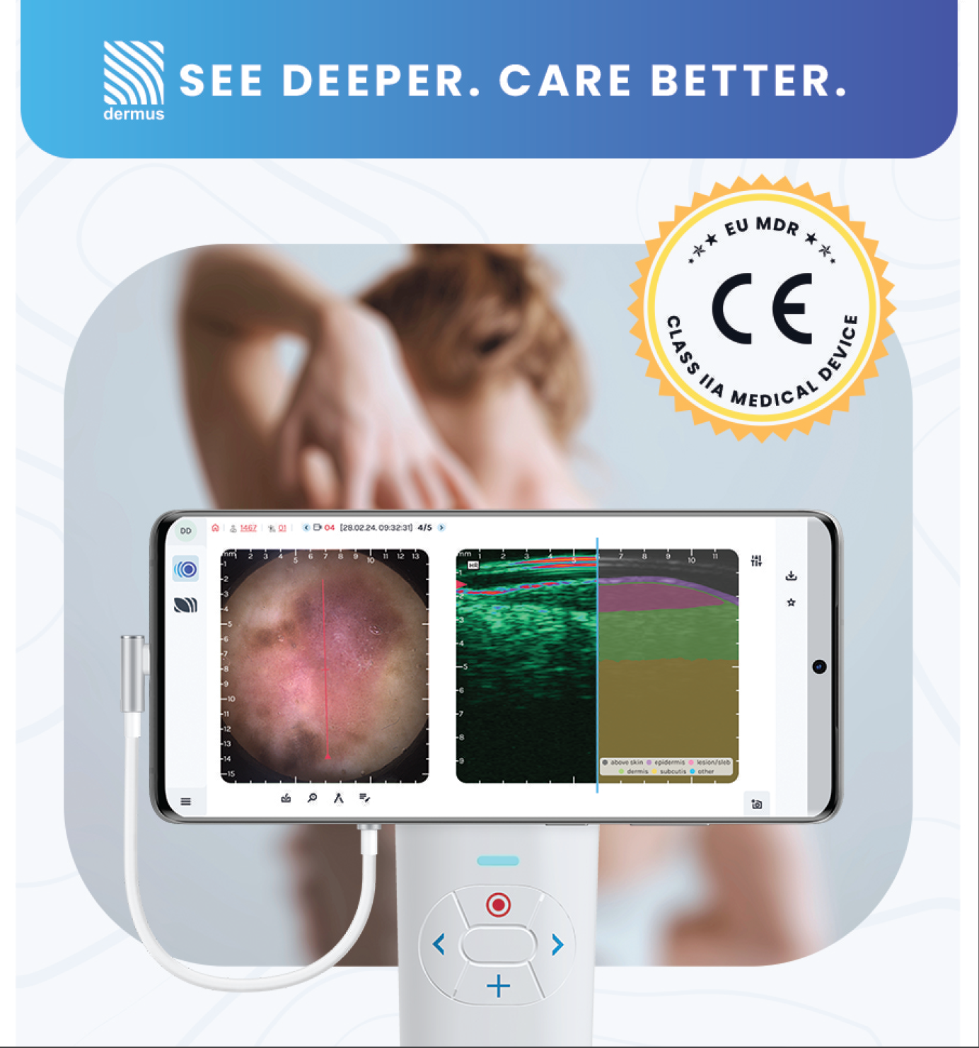
Handheld high-frequency ultrasound for skin (20-40 MHz, down to 10 mm) with a dermoscopy camera in the same head. Measures lesion and wound depth, thickness and volume, images blood flow with 2-D power Doppler, and returns to the same spot at every time point. CE Class IIa under EU MDR, FDA pending. Published preclinical work in mouse skin tumor, UV inflammation and wound healing models.


Overview
SkinScanner is a handheld ultrasound for skin. The probe runs at 20-40 MHz and reaches 10 mm deep.
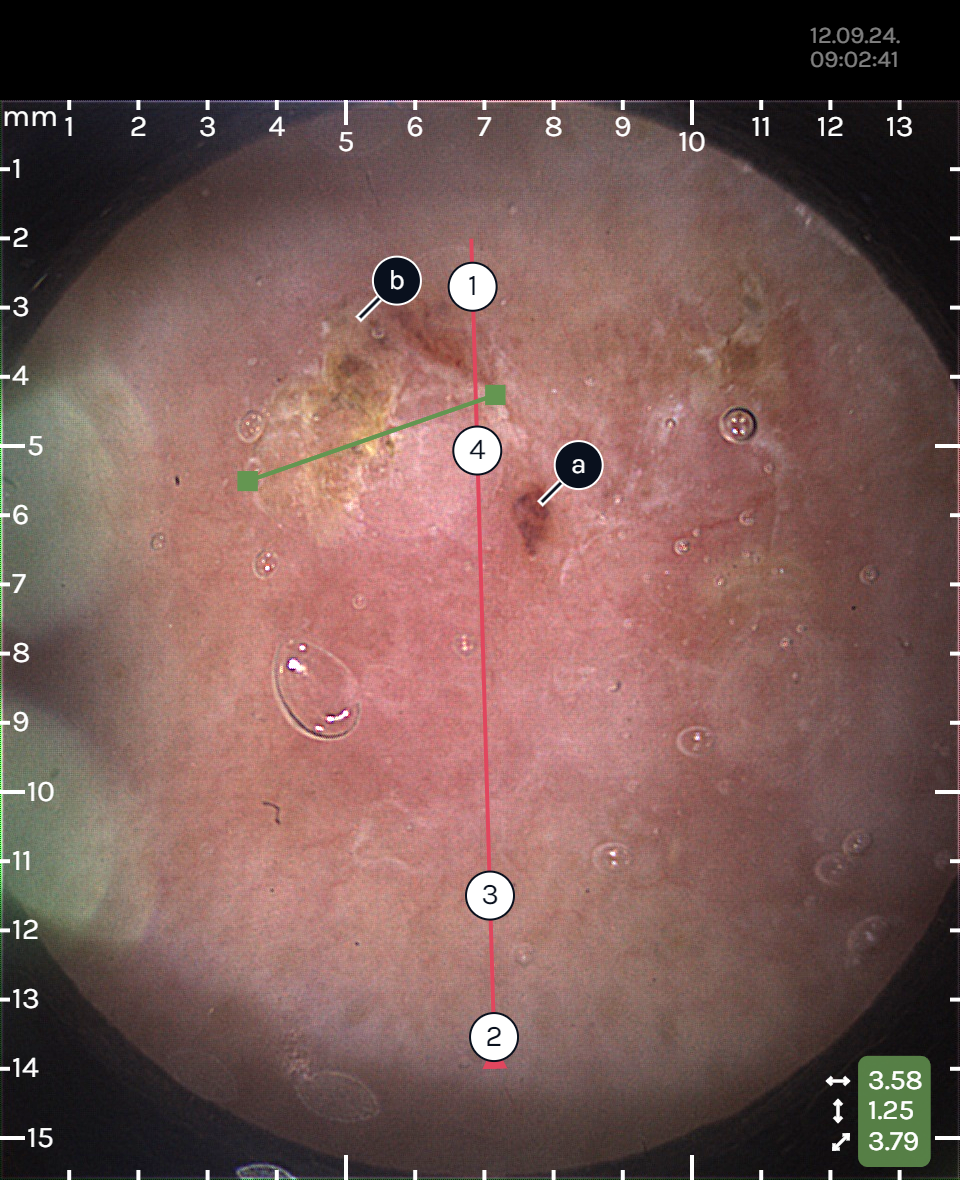
A dermoscopy camera in the same head records the surface at the same moment. A positioning line on the dermoscopy image marks where the ultrasound slice cuts through it.
The positioning line is what makes follow-up scans comparable. Without it, a handheld scan on day 3 and another on day 28 are two different slices.
CE Class IIa under EU MDR for clinical use in the EU. FDA application pending.
Imaging specifications
- Frequency: 20-40 MHz
- Depth: up to 10 mm
- Ultrasound field of view: 10 mm (depth) x 12 mm (lateral)
- Frame rate: 4+ fps
- Resolution: 100 µm axial, 300 µm lateral
- Optical field of view: 12 x 12 mm
What it measures
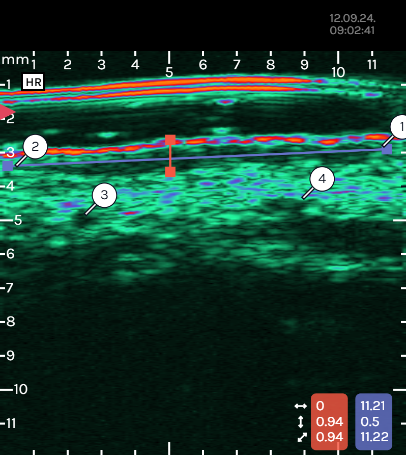
- Geometry: 2-point distance (thickness, diameter), AI-assisted segmentation of the skin layers, volume estimation.
- Intensity: echogenicity of a region over time, used to follow hydration, collagen and vascularization trends.
- Doppler: 2-D power Doppler for blood flow imaging.
Analysis runs in SkinAid, the Dermus web-based software.
Where it is used
- Skin cancer: depth and extent of melanoma, BCC and SCC before surgery.
- Inflammation: psoriasis, hidradenitis suppurativa, rosacea, acne.
- Aesthetics: filler placement and the vessels under it.
- Preclinical: mouse skin tumor, UV inflammation and wound healing models. Lesion and wound depth, thickness and volume in the same animal at every time point.
Where it is not
Anything deeper than 10 mm. Orthotopic tumors in internal organs, cardiac and abdominal imaging require a full preclinical ultrasound system.




Dermatology
צפו בכל המוצריםDermus Publications
Dermus high-resolution ultrasound skin imaging systems enable non-invasive assessment of skin structure and treatment response. They are used in dermatology research, aesthetic medicine, and clinical studies evaluating skin thickness, elasticity, and the efficacy of topical and laser-based interventions.
Nonthermal atmospheric pressure plasma (NTAPP) effectively eliminates skin tumors in a mouse model of UV-induced carcinogenesis with a short-term treatment regimen
Skin cancer model in SKH1 hairless mice. UV-induced skin tumors treated with a one-week nonthermal plasma protocol.
Acute UV response of early erythema and late edema in SKH1 mice
Skin inflammation model in SKH1 mice. Early erythema and late edema after acute UV exposure.
Hybrid Nanofibers for Multimodal Accelerated Wound Healing
Wound healing in mice under drug-loaded hybrid nanofiber dressings, followed to day 11.
What does the SkinScanner measure?
Skin and skin lesions in depth. A handheld high-frequency ultrasound (20-40 MHz, down to 10 mm) works together with a built-in dermoscopy camera that records the surface at the same moment. Outputs include thickness, depth, diameter and volume of a lesion, segmentation of the skin layers, and blood flow with 2-D power Doppler.
Can it be used on mice?
Yes. Published preclinical work with the SkinScanner covers UV-induced skin tumors in SKH1 mice, acute UV inflammation (erythema and edema) in SKH1 mice, and wound healing under nanofiber dressings in mice. The same animal is scanned at every time point, with no group sacrificed for histology at each one.
How is the same spot found again on day 3, day 7 and day 28?
The dermoscopy image (12 x 12 mm) is recorded together with the ultrasound frame, and a positioning line marks where the ultrasound slice sits on the surface. Matching the surface image to the previous session puts the probe back on the same slice.
Is the SkinScanner available in Israel with local support?
Yes. NBT is the exclusive distributor in Israel and handles installation, application training and service.
When is the SkinScanner the wrong tool?
When the target sits deeper than 10 mm, for example orthotopic tumors in internal organs or cardiac imaging. Those require a full preclinical ultrasound system. Skin lesions, wounds and subcutaneous tumors within 10 mm of the surface are within range.
יש לכם שאלות נוספות?
בקשה להצעת מחיר
אנא מלאו כמה שיותר פרטים ואנו נטפל בבקשתכם בהקדם האפשרי
בקשה להצעת מחיר
אנא מלאו כמה שיותר פרטים ואנו נטפל בבקשתכם בהקדם האפשרי