.svg)
Micropatterning 101: Turning Cell Geometry Into an Experimental Variable
Cells cultured under identical conditions still behave differently, and part of that variability is simply spatial randomness. This ibidi primer explains how micropatterning turns geometry into a controlled experimental parameter.
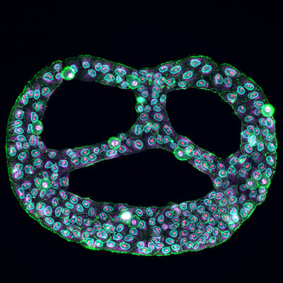
Anyone who has imaged a standard culture dish knows the problem: some cells spread out broadly, others stay compact; some polarise and migrate, others divide in place. We tend to file this away as biological heterogeneity, and part of it genuinely is. But a substantial share of it is something more mundane - spatial randomness. Where a cell happens to land, how much surface it can grab, and which neighbours it touches all feed directly into signalling, migration speed, differentiation, drug response and survival.
A new primer on the ibidi blog, written by Abhishek Derle, PhD, makes the case that micropatterning addresses this at the root.
What micropatterning actually does
Micropatterning defines precisely where cells are allowed to attach. Instead of one large adhesive surface, the substrate carries small adhesive islands on a passivated background - the areas outside the pattern are engineered to resist both cell attachment and protein binding, so adhesion is confined to the intended shapes.
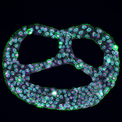
Those islands can be circular, square, linear or fully custom, and they are defined at micrometre scale. By tuning pattern size and spacing, you control how many cells land per spot, how far apart neighbours sit, and whether cells stay isolated or assemble into clusters. Geometry stops being an uncontrolled variable and becomes something you set deliberately.
The technique itself is not new - early work relied on stamping ECM proteins onto surfaces with elastomer moulds, which worked but demanded real technical skill. Improvements in surface chemistry made background passivation and pattern stability far more reliable, and ready-to-use micropatterned labware has since removed the fabrication step entirely.
Four geometries, four kinds of question
- Single-cell arrays. Small adhesive regions with defined spacing let individual cells attach in isolation. This matters most in imaging-based assays, where overlapping cells wreck segmentation and quantification. Spatial separation alone can meaningfully improve data quality in morphological or fluorescence-intensity work.
- Multi-cell arrays. Larger adhesive areas let several cells settle within one defined region, forming reproducible micro-colonies. Useful for cell-to-cell communication studies, tumour microenvironment models and immune cell interaction assays. Defined colony geometries have even been used to reproduce aspects of early embryonic spatial organisation in vitro.
- Spheroid formation. Seeding cells in suspension onto adhesive spots surrounded by a fully non-adhesive background produces compact, uniform aggregates, which cuts the spheroid-to-spheroid variability that plagues screening and drug testing.
- Line arrays. Linear patterns create micro-lanes that guide orientation and directional growth. Valuable for neurite extension, cytoskeletal organisation and structured migration studies; one-dimensional confinement has been shown to mimic fibrillar in vivo migration better than unconstrained 2D culture.
Adding flow to the picture
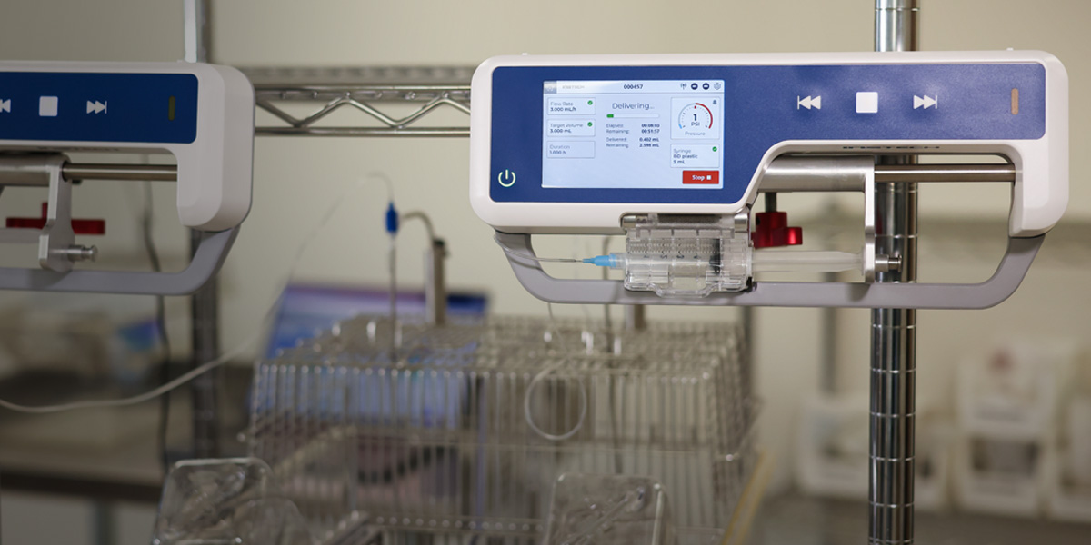
Tissue in vivo is rarely static. Micropatterning can be combined with perfusion so that patterned cells or aggregates sit in a perfusable slide system under continuous media circulation and defined shear stress. Structured spheroids held under flow can show improved compactness and long-term stability, while adherent cells get mechanical stimulation without losing their spatial arrangement. Combining geometric confinement with fluid dynamics moves the experiment considerably closer to tissue-like conditions.
Why it matters now
Modern cell biology leans heavily on imaging, quantification and reproducibility. High-content analysis, AI-based image processing and spatial omics all benefit from standardised cell organisation. Micropatterning reduces spatial noise and improves comparability across wells, time points and laboratories - defined target-cell arrays simplify live interaction assays in immunology, uniform spheroids support reproducible screening in toxicology and drug development, and guided micro-lanes clarify directional growth in neuroscience.
The framing in the original article is worth keeping: micropatterning is not just a way to constrain where cells attach. It is part of experimental design - a way to build spatial control into the logic of the study rather than adapting the study to whatever the dish happens to produce.
NBT supplies the full ibidi range in Israel, including standard and custom micropatterned labware, the ibidi Pump System and stage top incubators. Get in touch if you would like to discuss which pattern geometry fits your assay.
Source: ibidi blog.

NEED MORE INFORMATION ABOUT THIS PRODUCT?
Send us your emailRecent Blog Posts

.svg)

Advance Your Research
Contact NBT today for expert consultation on your neuroscience instrumentation needs.