.svg)
.svg)
ibidi µ-Slide 8 Well high µ-Pattern
Eight well slide with a micropatterned adhesive surface, in four pattern geometries: squ30 pit75 hex (83811), cir200 pit600 hex (83812), cir500 pit1000 hex (83813) and lin20 pit170 (83814). Tell us which pattern the protocol calls for and we quote that one.

What is the µ-Slide 8 Well high µ-Pattern?
A specialized micropatterned slide combining eight wells with defined cell-attachment patterns on the coverslip surface. The µ-Pattern technology provides micro-scale geometric cues to control cell morphology, migration, and organization. Available in multiple pattern types (circular, hexagonal, and pit designs with varying pitch), this slide enables quantitative studies of cell behavior on patterned substrates.
The µ-Slide 8 Well high µ-Pattern bridges high-throughput imaging with geometric cell guidance, ideal for developmental biology, cell migration, and mechanotransduction research.
Key Features
- Micropatterned surface – Defined cell-attachment regions guide cell organization
- Multiple pattern geometries – Circular, hexagonal, and pit designs available
- 8-well format – Each well with consistent patterned substrate
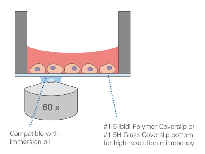
- High optical clarity – #1.5 Polymer or Glass Coverslip bottom
- Quantitative analysis – Precise cell positioning for reproducible measurements
.webp)

Technical Specifications
| Catalog Numbers (Consolidated) | IBD-80838 (cir200, pit600, hex), IBD-80840 (cir500, pit1000, hex), IBD-80843 (cir500, pit1000, hex - ibiTreat), IBD-80850 (custom patterns) |
| Number of Wells | 8 |
| Well Volume | 300 µL |
| Pattern Types | Circular (cir200, cir500), Hexagonal (hex), Pit (pit600, pit1000) |
| Surface Coating | ibiTreat or Untreated |
| Coverslip Bottom | #1.5 Polymer or #1.5H Glass |
| Slide Format | ISO 8037/1 standard microscope slide |
| Packing | 5 per box |
Applications
- Cell migration and wound healing assays
- Morphology and organization studies
- Mechanotransduction research
- Developmental biology and pattern formation
- Quantitative live-cell and fixed imaging

Fluorescence Imaging
Explore All ProductsCell Cultivation and Imaging
Explore All Productsibidi Labware Publications
Comprehensive collection of peer-reviewed research publications demonstrating the use of ibidi labware including µ-Slides, µ-Dishes, µ-Plates, and specialized microfluidic devices for cell biology, neurobiology, vascular biology, and cancer research.
What is it used for?
Micropatterned eight-well slide for cell guidance and morphology studies
Is it compatible with high-resolution microscopy?
Yes. The #1.5 ibidi Polymer Coverslip bottom has low autofluorescence and low birefringence, and it handles widefield fluorescence, confocal, phase contrast and DIC. For TIRF, single molecule detection and super-resolution, buy the Glass Bottom version instead.
Is it sterile and ready to use?
Yes. Sterilized and individually wrapped
Is local support available in Israel?
Yes. NBT is the ibidi distributor in Israel
Is it suitable for live-cell imaging?
Yes. Pair it with an ibidi Stage Top Incubator for CO2 and temperature control and it will run long term live cell imaging.
Still have questions?
Related Products:
Request for Quote
Please fill in as much details as possible and we will take care of your request as soon as possible
Request for Quote
Please fill in as much details as possible and we will take care of your request as soon as possible