.svg)
Smarter Imaging: AI Meets Automation in Cell Microscopy
Automation and AI are transforming cell microscopy — from robotic stages and standardized acquisition to deep learning-powered analysis of 3D organoids, enabling reproducible, quantitative imaging at scale.
How Automation Transformed Cell Imaging
The integration of automation and artificial intelligence has reshaped cell imaging. Automation arrived first, changing the day-to-day reality of imaging by automating repetitive work and standardizing acquisition on a large scale. Robotic stages keep hundreds of wells in reliable focus. Acquisition software applies consistent exposure and illumination from plate to plate. Incubation systems maintain a steady temperature and CO₂ level, allowing live cells to behave naturally.
The result is clean, repeatable images with far less hands-on effort and far fewer sources of variability.
AI as the Next Layer
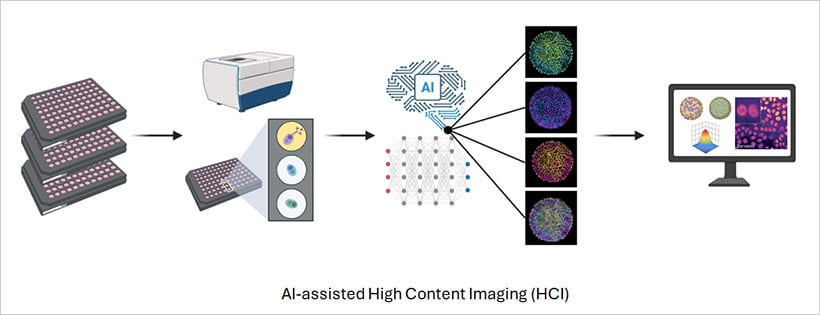
Artificial intelligence became the next layer on top of automation. It turns large image sets into quantitative answers by detecting cells and organelles, and measuring intensity, texture, shape, and movement across channels and time. Recent advances in machine learning — especially deep learning — enable models to find subtle, hidden structures in both simple and complex imaging data.
Deep learning models learn phenotypes directly from data, enabling tasks like virtual staining and label-free imaging that were previously impossible. Together, automation and AI are increasing reproducibility across plates, instruments, and sites. This consistency is essential for large-scale, multi-site experiments, helping to turn imaging into a truly quantitative discipline.
Compatibility with 3D Models
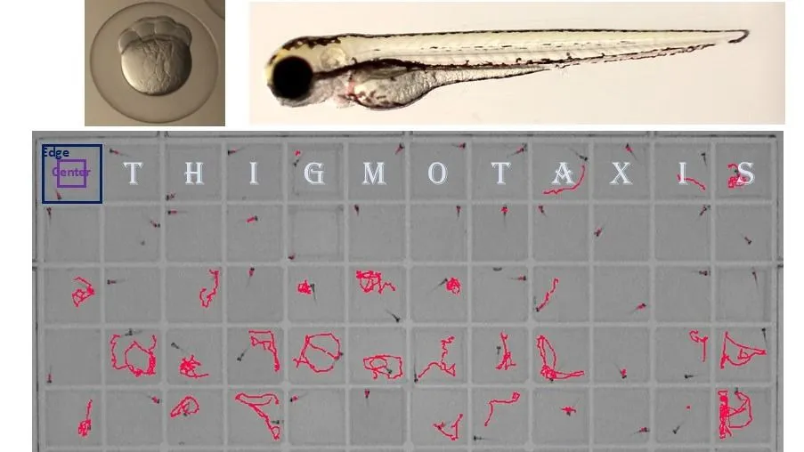
As biology advances toward more realistic models, AI and automation enable the study of 3D spheroids, organoids, and co-cultures with the same precision as 2D systems. Automated microscopes maintain focus through thick samples, while AI algorithms reconstruct and quantify 3D structures over time.
In patient-derived organoid studies, high-content imaging combined with AI has already helped predict how individual tumors respond to specific drugs, paving the way for data-driven precision medicine. The ibidi µ-Plate 96 Well 3D supports uniform spheroid formation and stable imaging over long time courses, providing a reliable platform for quantitative high-content imaging of complex models.
What This Means for Cell Biology Research
The combination of automation and AI is not just a technical upgrade — it represents a shift in what is experimentally possible. Screens that once required weeks of manual effort can now be completed in days, with more consistent results and richer datasets. Researchers can ask more complex questions, work with more biologically relevant models, and generate data that scales with the demands of modern science.
For labs working with ibidi labware, these advances integrate directly into existing imaging workflows, enabling high-content screening, live cell analysis, and 3D model studies with minimal adaptation.
NEED MORE INFORMATION ABOUT THIS PRODUCT?
Send us your emailRecent Blog Posts


Advance Your Research
Contact NBT today for expert consultation on your neuroscience instrumentation needs.