.svg)
Organoids or Organ-on-a-Chip? What Each Model Actually Gives You
Organoids self-organise but lack vasculature and drift over time. Chips give you perfusion and mechanical control but are laborious to fabricate. Elveflow compares the two model classes honestly, and makes the case for combining them.
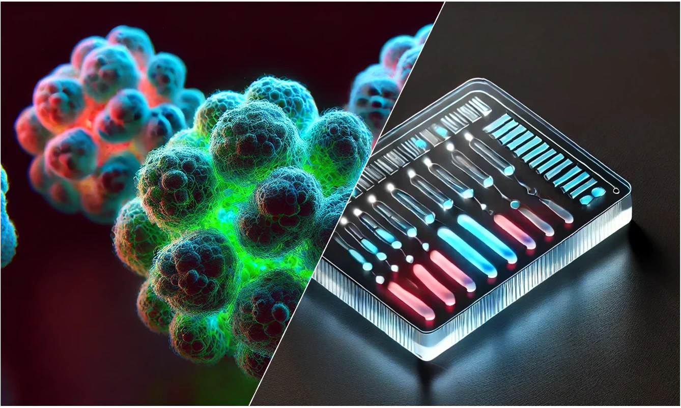
Microphysiological systems get discussed as one category, which obscures a real difference. Organoids and organ-on-a-chip devices solve different problems, fail in different ways, and suit different questions. A comparative review by Louise Fournier, PhD on the Elveflow site lays the two side by side, and it is a useful piece to have to hand when someone in the lab asks which one they should be building.
Organoids: self-organisation
Organoids are three-dimensional cellular structures that mimic the architecture and function of human organs. The lineage traces to 2009, when Hans Clevers' group showed that a single LGR5+ intestinal stem cell could differentiate and form an entire crypt-villus structure in 3D culture - cells embedded in Matrigel with R-spondin 1, EGF and BMP inhibitors. Models have since been established from nearly every major human tissue: brain, lung, liver, pancreas, kidney.
The mechanism is the selling point. Development relies on the self-organising capacity of adult stem cells or iPSCs, grown in a gel-like matrix that mimics the extracellular matrix, with organ-specific media formulations driving differentiation. What emerges are tissue-specific features (epithelial layers, glandular structures, neuronal networks) that nobody had to engineer directly.
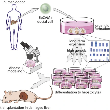
That makes them strong in cancer biology, where tumour organoids retain the genetic and histological features of the original tumour; in personalised medicine, where patient-derived cells give insight into individual drug response; in infectious disease modelling; and in developmental biology, where the point is precisely to watch organogenesis happen.
Where they run out
The limitations are structural rather than incidental. Organoids typically lack immune, nervous and circulatory systems, which rules out a whole class of cross-system questions. Without vascularisation, interior cells can be starved of nutrients and oxygen, producing necrotic cores. Reliance on gels like Matrigel brings batch-to-batch variability and limited environmental control. Genetic drift can alter the cell population over extended culture. And tissue-specific media formulations add cost and complexity.
Organ-on-a-chip: engineering
Organ-on-a-chip takes the opposite route. Microfluidic devices are designed to reproduce tissue-tissue interfaces, mechanical forces and chemical gradients, housing different cell types together to mimic the interplay found in living tissue.
Typically built from optically transparent materials such as PDMS, these systems feature microchannels lined with living cells, often separated by semipermeable membranes or embedded in ECM gels, under controlled flow. That flow does real work: efficient transport of nutrients and waste, mechanical stimulation, and precise management of the microenvironment including defined chemical gradients.
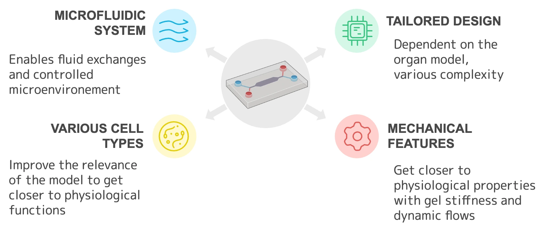
The applications follow from the control: high-throughput drug screening with efficacy and toxicity assessed in a defined environment, disease modelling (lung-on-a-chip for pulmonary disease and inhaled therapeutics), hepatotoxicity testing, multi-organ "body-on-a-chip" platforms for systemic effects, and reduced reliance on animal models.
Where they run out
The costs are practical. These systems require careful design and precise operation, with real setup and maintenance overhead. Many labs lack access to soft lithography and microfabrication, which is driving interest in laser cutting, adhesive film stacking and 3D printing as alternatives. Connecting devices to pumps and tubing raises contamination risk. Bubbles in microchannels obstruct flow and compromise viability. Producing consistent, reproducible chips is labour-intensive and costly. And standardisation and regulatory approval remain open problems for wider adoption.
Organoids-on-a-chip
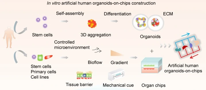
The interesting part of the review is the convergence. Integrating organoids into microfluidic platforms gives the self-organised tissue a controlled perfusion environment, mechanical stimuli and biochemical gradients - addressing the vascularisation and variability problems from the organoid side while keeping the biological complexity that engineered chips struggle to produce.
The review points to better modelling of organ-specific functions such as vascularised tissue, the ability to study organ-organ interaction dynamically, higher reproducibility and better scalability.
What has to happen next
Three priorities are flagged for the field: material research, particularly new culture media and ECM-mimicking hydrogels that supply the right structural and biochemical cues; deep learning and automated image analysis, to handle the datasets these systems generate and improve reproducibility; and global collaboration on standardisation, including validation criteria and regulatory frameworks, without which neither technology translates.
The honest summary is that these are complementary rather than competing. Organoids capture genetic and histological fidelity; chips supply a dynamic, perfused environment with controlled conditions. Which one you want depends on whether your question is about the tissue or about what the tissue is exposed to.
NBT supplies Elveflow microfluidics in Israel - the OB1 pressure controller, flow sensors, MUX valves and application packs for organ-on-a-chip, perfusion and recirculation - alongside the ibidi range for 3D culture and imaging. Get in touch to discuss flow control for your model.
Source: Elveflow Microfluidic Reviews.
NEED MORE INFORMATION ABOUT THIS PRODUCT?
Send us your emailRecent Blog Posts

.svg)

Advance Your Research
Contact NBT today for expert consultation on your neuroscience instrumentation needs.