.svg)
Musculoskeletal Modeling
Electromyography-driven musculoskeletal models can track muscle fatigue in real-time, with applications in sports training, rehabilitation, and manual handling. This post explores a novel time-varying model (t-pEMS) that uses EMG data to estimate fatigue progression with person-specific accuracy.
Key Takeaways
- Musculoskeletal modeling uses muscle behavior to create a computational model that can understand muscle fatigue and risk of injury. Personalized EMG-driven musculoskeletal models (pEMS) take in EMG signals from a patient to determine fatigue changes.
- Novel t-pEMS model: a pEMS coupled with muscle force decay models that tracks person-specific fatigue progression and accurate fatigue estimation from joint muscle-moments during functional tasks.
- The t-pEMS model was able to characterize fatigue while being patient specific — applicable in sports training, rehabilitation of post-musculoskeletal disorders, or manual handling in factories.
What is Musculoskeletal Modeling?
Musculoskeletal modeling is the computational modeling of the musculoskeletal system to determine and study muscle behavior, such as muscle kinematics and muscle fatigue. Modeling muscle behavior can provide insight into incidence and progression of fatigue. Specifically, understanding muscle fatigue is needed to discern muscle overuse and risk of injury that leads to muscle disorders.
Personalized electromyography-driven musculoskeletal models (pEMS) are a type of musculoskeletal model that is human-specific and utilizes EMG to determine fatigue changes. These models can provide insight into the influence the patient has on the outcome of the model.
Fatigue-Based Musculoskeletal Modeling
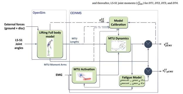
Effective musculoskeletal fatigue modeling simulates both the central and peripheral components of fatigue. To do this, changes in neural drive or EMG activity and their influence on the model need to be measured. Most current fatigue models are not capable of capturing both components of fatigue, thus increasing inaccuracy. By utilizing EMG activity, pEMS coupled with muscle force decay models results in a model called time-varying pEMS (t-pEMS) that captures both components.
How EMG Was Used
Delsys EMG sensors were used to measure EMG that contributed to the calibration of the pEMS model. EMG signals were mapped in the LFB model according to the muscle-tendon units (MTUs), and EMG amplitude was used in analyzing the functional tasks. In the t-pEMS, EMG was utilized in the modeling of the maximum isometric force and peak contraction velocity as the measure of muscle-specific history.
What They Found
The t-pEMS reduced overestimation of body-weight lumbosacral moments (BW-LM) compared to the pEMS alone. The influence of fatigue varies among different muscles and individuals, as EMG activity naturally varies across individuals and muscles. Muscle activity increased as functional tasks proceeded due to increasing fatigue, but EMG amplitude did not change consistently during each subsequent task.
Future Impact
This model can be applied to a vast range of applications where fatigue presents as an issue: sports training, rehabilitation post-musculoskeletal disorders, or manual handling in factories. The person-specificity aspect makes the model more robust and reliable. EMG allowed researchers to access and incorporate fatigue of the muscles into the model, making it more accurate and applicable to real-world activities.

Shall we chat about this topic?
Contact usRecent Blog Posts


Advance Your Research
Contact NBT today for expert consultation on your neuroscience instrumentation needs.