.svg)
When Microstructure Meets Mapping: What a New DES Study Reveals About Language Networks
A new Clinical Neurophysiology study shows that cortical microstructure — specifically myelin content — shapes the earliest components of direct electrical stimulation responses, with implications for intra-operative brain mapping and why the g.HIamp amplifier’s artifact control matters.
A recent publication in Clinical Neurophysiology by Turpin et al. (2025) provides new insight into a question neurosurgeons and neuroscientists encounter daily in the operating room: why does the brain respond so differently to the same electrical stimulation, depending on where you stimulate?
The answer lies deep in cortical microstructure — and it highlights why high-fidelity biosignal amplifiers such as the g.HIamp are becoming indispensable for both clinical research and surgical decision-making.
The Study
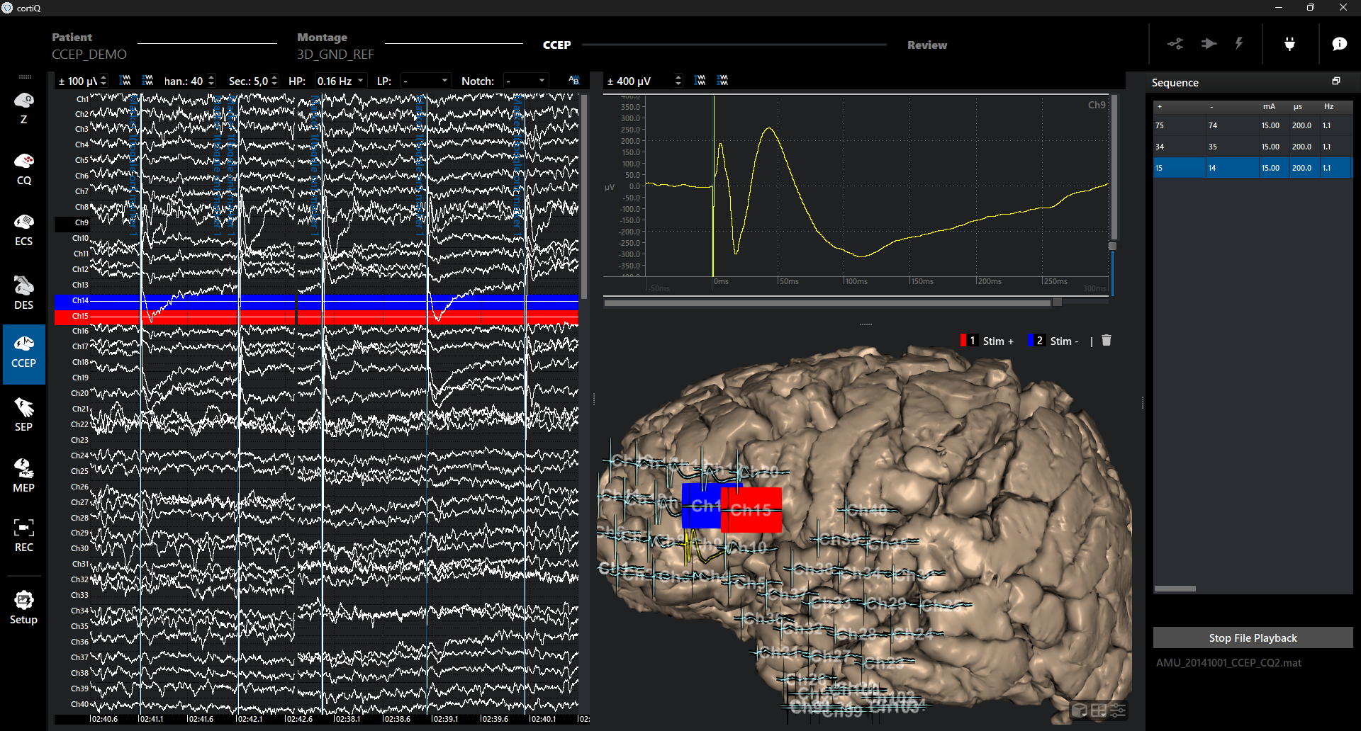
The authors investigated direct cortical responses (DCRs) evoked by direct electrical stimulation (DES) during awake brain surgery. In 10 patients undergoing tumor resections, DES was applied to different cortical regions while electrocorticography (ECoG) signals were recorded using the g.HIamp amplifier.
The analysis focused on the earliest evoked response components: the P0 (fast positive component within 10ms, related to action potentials) and N1 (negative deflection within 30ms, related to postsynaptic potentials).
Key Finding: Myelin Matters
Motor (M1) and primary sensory cortex (S1) show a significantly steeper P0 slope than associative regions such as Broca’s or Wernicke’s areas. This directly reflects myelo-architecture: primary cortices contain larger, heavily myelinated fibers that conduct electrical activity faster and more synchronously.
In simple terms: the very first milliseconds of the evoked response already encode information about the tissue’s wiring. This reinforces a growing view that early evoked components are not just artifacts of stimulation — they are biomarkers of cortical structure.
Why Signal Quality Is Critical
Detecting differences in the slope of the P0 component requires very high temporal precision and minimal stimulation artifacts. The g.HIamp biosignal amplifier addresses this with DC-coupled, high-dynamic-range signal acquisition that allows the initial slope of P0 to be resolved rather than masked or clipped by residual artifacts.
Implications for Brain Mapping
Early DES-evoked components may one day help identify cortical areas even when task-based mapping is limited — for example, in time-critical surgical phases, under general anesthesia, or in pediatric patients. Building on these capabilities, cortiQ 2.0 adds dedicated stimulation hardware and switching to provide an integrated platform for clinical brain mapping and CCEP research.

Shall we chat about this topic?
Contact usRecent Blog Posts


Advance Your Research
Contact NBT today for expert consultation on your neuroscience instrumentation needs.