.svg)
Why Use Mesh MEA Instead of Planar MEA for Organoid Work?
As organoid research grows, traditional 2D planar MEAs show clear limitations. Mesh MEA from Multi Channel Systems maintains structural integrity, enables long-term recordings from inside intact 3D organoids, and reduces experimental risk.
As organoid research continues to grow and evolve, the limitations of traditional 2D planar microelectrode arrays (MEA) have given rise to the need for more advanced technologies. For researchers recording electrophysiological data from inside an intact organoid, Mesh MEA from Multi Channel Systems offers significant advantages over 2D counterparts.
The Core Problem with Planar MEA
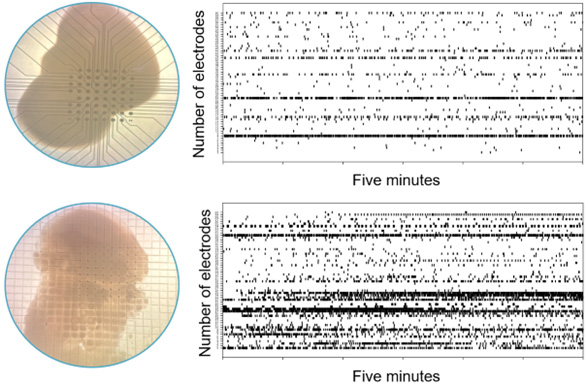
Traditional 2D planar MEA chips are ideal for cell cultures, stem cells, and tissue slices — but an organoid cultured on a 2D MEA chip will collapse and flatten. This may compromise the physiological responses of the organoid and impact the validity of the data. Mesh MEA allows the organoid to grow around a layer of mesh, maintaining its 3D shape while electrophysiological data is recorded from inside.
Structural Integrity and Data Quality
One of the primary advantages of Mesh MEA is its ability to maintain the structural integrity of organoids. Traditional 2D MEAs require the organoid to be transferred to the MEA platform, a process that often results in structural alterations. Mesh MEA allows long-term culture directly in the MEA chip, preventing deformation and preserving the organoid’s morphological structure.
Experiment Duration and Long-Term Measurements
Organoids placed on 2D MEAs tend to deform within a few days, jeopardizing long-term recordings. Mesh MEAs, with their structural integrity and integrated perfusion systems, allow for extended monitoring periods. This is crucial for chronic studies requiring repeated long-term measurements of cellular electrophysiology and drug effects over time.
Organoid Accessibility and Electrode Incorporation
Traditional 2D MEAs fall short in recording data from within organoids without causing structural damage. Mesh MEAs facilitate cellular migration around the electrode-containing mesh, allowing data recordings from the interior of the organoid — providing a more accurate representation of electrophysiological activities occurring within the 3D tissue structure.
Reducing Costs and Experimental Risks
Mesh MEAs enable the generation, maturation, maintenance, and measurement of organoids within a single instrument — reducing the likelihood of costly mistakes and spreading expenditures more effectively for long-term research projects.

Shall we chat about this topic?
Contact usRecent Blog Posts


Advance Your Research
Contact NBT today for expert consultation on your neuroscience instrumentation needs.